Color-Coded Cell Collection to Serve as Roadmap to Diseases
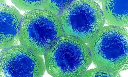
The Allen Institute for Cell Science has developed lines of color-coded stem cells that will serve as a powerful roadmap to our understanding of human disease. The cell collection consists of iPS (induced pluripotent stem cells) into which fluorescent tags have been inserted to target key structures within the cell with a clarity that has never been achieved before. This will allow scientists to visualize how healthy cells differ from diseased cells.
Executive director Rick Horwitz, Ph.D., explains: “Each cell is like a city. The color-coded cell lines can be likened to an online traffic map for the scientific community, enabling us to clearly and consistently see the cell’s dynamic organization. In other words, scientists can see which part of the cell is where at which time.”
The Technology
The scientists have used a technology known as CRISPR/Cas9 to fluorescently tag the major cell structures in human iPS. These tags are far more precise than current methods which involve indiscriminately flooding a fluorescent protein into the cell. The new technology allows precise targeting of key cellular structures which can then be tracked as the cell goes through various stages of its life cycle.
Pluripotent stem cells can be converted to a variety of cell types such as brain, heart, and skin. According to Anthony Hyman, Ph.D., these color-coded gene-edited lines will make iPS cells available to all cell biologists, thus lowering the entry threshold and ushering in a new era of innovation in cell biology.
The First Collection
The first collection from the Allen Institute for Cell Science includes five lines that target five main structures in iPS cells: the nucleus, mitochondria, cell junctions, microtubules, and adhesions. Further collections are expected in 2017 and will also be distributed through the Coriell Institute for Medical Research. These precisely tagged cell lines closely mimic normal human cells, allowing scientists to study them in the laboratory, test drugs on them, and determine how they differentiate, says Ruwanthi Gunawardane, Ph.D.
The Future
With these cell lines, scientists will be able to observe healthy cells in a manner that is unprecedented. “We will be able to see the cell’s structures with a clarity that is nothing short of astonishing,” says Susanne Rafelski, Ph.D. “This will give us a multi-structure broad overview of the changes that occur in a cell when it goes from a healthy state to a diseased state.”
The fluorescently tagged cell lines will be easily accessible to cell biologists globally and will help deepen our understanding of fundamental cell biology and human disease. For example, critical mutations can be induced in the cells and the behavior of the tagged cellular structures observed in a Petri dish. This could have a dramatic impact on the field of personalized medicine, where each therapy is tested and tailored to the individual patient rather than simply hoping the standard therapy will work.
References:
1. https://www.sciencedaily.com/releases/2016/11/161130155421.htm